2.4.4 - Calibration for Alpha Beta Discrimination (Triathler)
Introduction
As described in chapter 2.1.3. (fig. 13), α-nuclides appear at the same energy position as medium and high energetic β-emitters. This fact together with the poor energy resolution of 0.3 to 0.5 MeV prevents the distinction between α- from β-emitters. The Triathler system from HIDEX, which is described here as example, applies the Pulse Length Index (PLI) as α/β-discriminator. α-pulses with longer pulse length (duration) are measured in the upper α-channel, while other pulses below the PLI level are collected in the β-channel. The PLI level may individually be set in order to obtain optimized separation results.

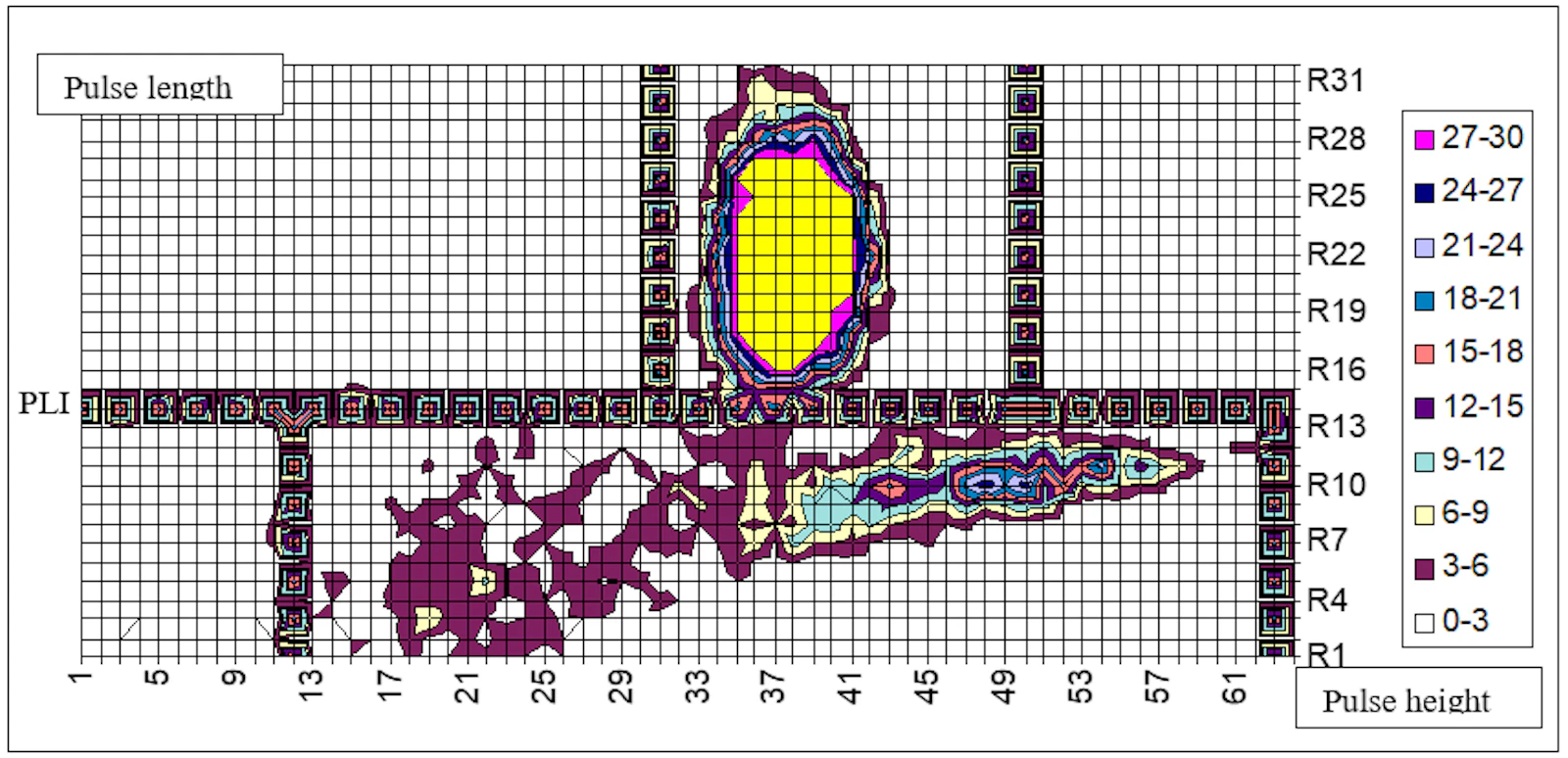
In the 2D PSD surface plot (e.g. fig. 15, 17) the energy (pulse height) on x-axis is plotted against the pulse duration (pulse length) on y-axis and the intensity as background color layout respectively.
The horizontal line as „Pulse Length Index“ PLI separates a- from b-pulses. a-pulses with longer pulse duration (upper part) are compiled in the a-channel, while the shorter β-pulses (lower part) are sorted in the β-channel.
The vertical lines correspond to the energy windows for α- (upper part) and b- (lower part) pulses. PLI and energy windows may be set individually in order to optimize α/β-separation and background. A three dimensional representation with the Triathler software can be created automatically by Excel Macros (fig. 16). With its outstanding α/β-separation capacity, the mobile Triathler device may be used for in-situ measurement of Radon in air and water (2.2.1.2. and 2.2.1.3.), as well as for 226Ra/228Ra (2.2.1.4.) and 238U/234U (2.2.1.8.) isotopes in water samples. From the Uranium isotope ratio, the origin of the sample may be identified e.g. as natural Uranium or as “Depleted Uranium” (DU). The mobile system is also suitable for in-situ contamination measurements using swipe assays (2.4.4.).

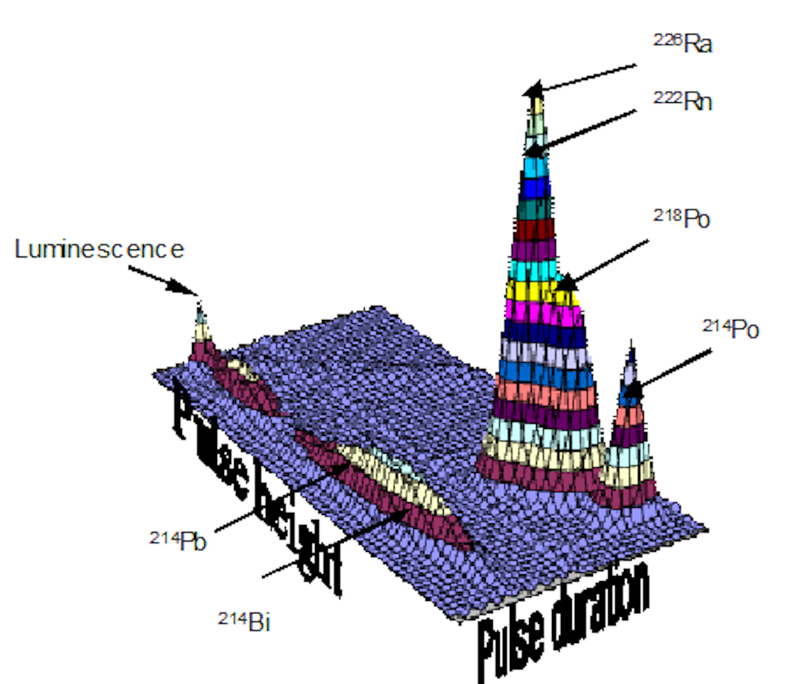
The representation of both the 2D surface plot and the 3D graphical presentation may be used for verifying a proper α/β-discrimination (fig. 18). Thus, interferences from β-emitters may be recognized immediately, estimated and corrected by proper setting of the PLI discriminator. It demonstrates an effective quality control of results by modern instrumentation.
Materials and Equipment
Standard solution of α- and β-emitting radionuclides
LS instrument with graphical α/β-PSD option (e.g. HIDEX Triathler, HIDEX SL300 or 600SL)
Procedure (for Triathler)
The program is defined in the extended mode of label 4 "alpha separation” and logarithmic scale.
A mixed α/β-sample is measured (if practicable 500 to 1000 counts) and the 2D plot visualized in the PC, automatically or after previous setting (3 enter). The 2D plot should appear in the middle part of the plot. The position may be changed by „Y-pos“ (higher value lowers the spectrum; for Y-gain, Bias V, Delay and Offset see Triathler-Manual).
PLI and energy window should be changed iteratively in order that a-pulses appear quantitatively above the PLI without allowing interferences from β-pulses.
It should be noted that in the former software version, the original 1024 x 1024 data set is compressed in our example (fig. 15) to 64 x 32 for visualization.
For example, in order to set the window value to 61 the upper level for α-pulses has to be set to 61 x 16 = 976. The data are transferred automatically after previous setting (α: 4 enter, β: 1 enter, and 2D: 3 enter).
Example:
Settings for organic phase (Rn)
A: 500 – 880 PLI: 510 Y-gain: 75 Y-pos: 48
Settings for aqueous gel phase (Rn)
A: 400 – 750 PLI: 490 Y-gain: 75 Y-pos: 45
